Explain What the Different Ekg Waveforms Represent
The PR interval represents the atrial depolarization and physiological delay of the stimulus in the atrioventricular AV node. The QRS complex one single heart beat corresponds to the depolarization of the right and left ventricles lower heart chambers.

P Wave Qrs Complex And T Wave Cardiac Physiology Nursing Notes Cardiac Nursing Cardiac Sonography
The electrical impulse then reaches the ventricles.

. The U-wave which is a positive wave after the T-wave appears occasionally on the ECG. Its normal value is between 012 s and 020 s. V4 5th intercostal space in the midclavicular line.
Between the waveforms are the following segments and intervals. The P wave is the first positive deflection on the ECG. P Q R S T corresponding to different phases of the heart activities.
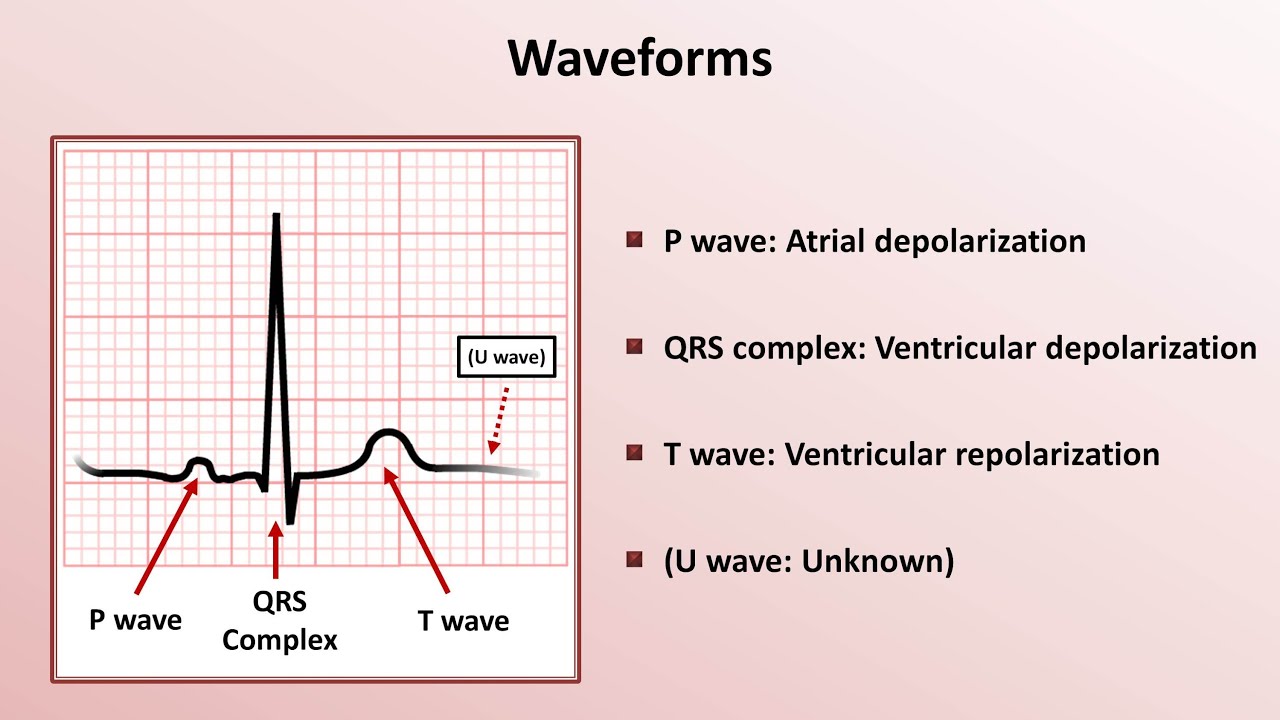
The P-wave represents the depolarisation of the atria which leads to the contraction of both the atria. ECG interpretation traditionally starts with an assessment of the P-wave. The P wave represents the spread of a stimulus through the atria atrial depolarization.
The PR interval is the distance between the onset of the P-wave to the onset of the QRS complex. V5 left anterior axillary line at the same horizontal level as. Duration of QRS complex is 006 to 010.
Duration of Q wave in the limb leads is 004 sec. Its the first negative waveform following the R wave. Together these waves represent one heartbeat.
It represents the time between two QRS complexes. Which wave represent Atrial depolarization. The different waves that comprise the ECG represent the sequence of depolarization and repolarization of the atria and ventricles.
Identify the Cardiac Axis. The three waves of the QRS complex represent ventricular depolarization. Start studying EKG waves segments intervals and complex.
A wave that is travelling towards the positive lead will inscribe an upwards deflection of the EKG. V3 midway between the V2 and V4 electrodes. The P wave represents the normal atrium upper heart chambers depolarization.
Learn vocabulary terms and more with flashcards games and other study tools. The T-wave represents ventricular re-polarisation. Represents late ventricular depolarization.
When this electrical impulse passes through your heart electrical currents are created that. If the wave immediately after the P wave is an upward deflection it is an R wave. The QRS complex represents stimulus spread through the ventricles ventricular depolarization.
Are the different options for a sensitivity button. There are three main components to an ECG. The P-wave PR interval and PR segment.
In a normal ECG theres three distinct waves. In order to determine if a rhythm is regular mark out several consecutive R-R intervals on a piece of paper then move them along the rhythm strip to check if the subsequent intervals are the same. Its height amplitude is approximately one fourth of the amplitude of the T-wave.
Conversely a wave traveling away from the positive lead will inscribe a downward deflection. ECG Waves and Intervals. Waves are the different upward or downward deflections represented on the EKG tracing.
The RR-interval starts at the peak of one R wave to the peak of the next R wave. 1 small box and 13 the height of the R wave in that lead. The ST segment and T wave represent the return of stimulated ventricular muscle to the resting state ventricular repolarization.
The standard 12-lead electrocardiogram is a representation of the hearts electrical activity recorded from electrodes on the body surface. They are the product of the action potentials created during the cardiac stimulation and repeated from one heart beat to another barring alterations. An ECG has three main components.
The PR interval is assessed in order to determine whether impulse conduction from the atria to the ventricles. As the ECG trace is recorded there are a series of upwards and downwards deflections created that represents atrial and ventricular depolarisation and repolarisation. What is the normal range for PR interval.
V2 4th intercostal space at the left sternal edge. Its the first positive waveform following P wave. Represents early ventricular depolarization.
The normal T-wave is somewhat asymmetric with a steeper downward slope. The hearts electrical activity is represented on the monitor or ECG tracing by three basic waveforms. This section describes the basic components of the ECG and the lead system used to record the ECG tracings.
Leads and their geometry. These are known as the ECG waves. V1 4th intercostal space at the right sternal edge.
In cases such as pre-excitation syndromes the PR interval may be shortened it. Definition Wave Types Electrocardiogram ECG. The P wave which denotes depolarising atria.
And the T wave represents repolarising ventricles. What does each wave of an ECG represent. The axis of an ECG is the major direction of the overall electrical activity of the heart.
And the T wave which represents the repolarization of the ventricles. Each ECG cycles consists of 5 waves. The ECG is recorded at a speed of 25 mmsec 5 large squaressec and the voltages are calibrated so that 1 mV 10 mm 2 large squares in the vertical direction.
If it is a downward deflection it is a Q wave. The QRS complex represents the depolarisation of the ventricles which leads to the ventricular contraction. There are six chest electrodes.
This can be seen in the Q R and S waves of the ECG which is called the QRS complex. The P wave the QRS complex and the T wave. The Standard 12 Lead ECG.
Then the T wave shows that the electrical impulse has stopped spreading and the ventricles relax once again. Shorten or lengthen the waves. Heart diseases and irregular heartbeats can be detected in ECGs.
The T wave represents the re-polarization or recovery of the. A U wave is sometimes present. The U-wave is most often seen in leads V2 V3 and V4.
The P-wave reflects atrial depolarization activation. For the inexperienced one of the most confusing aspects of ECG reading is the labeling of these waves. The PR interval the PR segment the ST segment and the QT interval.
The EKG waveform comes from a measurement of surface voltages between 2 leads. It is a small smooth-contoured wave and represents atrial depolarisation. The QRS complex which represents the depolarization of the ventricles.
It is seen as a small wave after the QRS complex in all leads except aVR and lead V1. It is measured from the beginning of the P wave until the beginning of the Q or R wave. There are mainly three types of ECG peak.
The P wave which represents the depolarization of the atria. Are the different. The QRS complex denotes the depolarization of the ventricles.
Intro To Ekg Interpretation Waveforms Segments And Intervals Youtube
A Typical Ecg Waveform And Its Characteristic Patterns P And T Waves Download Scientific Diagram

Associate Degree Nursing Physiology Review Cardiac Nursing Nursing School Survival Nursing School Studying
P A C S And The Heart And Extra P Wave Google Search Cardiac Nursing Nursing School Notes Nursing Notes


Comments
Post a Comment